S.M. Scientific Instruments (P) LTD

Home ≫Mortuary >Virtual Dissection Table and Digital Systems > Digital Dissection Table > Digital Virtual Anatomy Table SMI-DVAT

CLICK TO SCAN
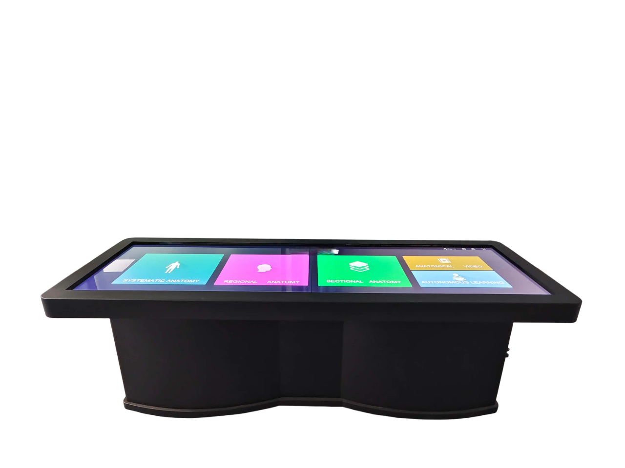
The Digital Virtual Anatomy Table categorizes anatomy into systematic anatomy, regional anatomy, and sectional anatomy and contains over 6,000 3D anatomical structures, enabling more detailed learning. It includes real dissection videos and anatomical animations to visualize the anatomical process. Additionally, it offers a vast collection of test questions to enhance knowledge consolidation.
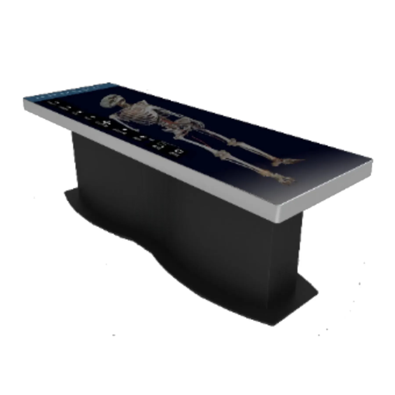
The Digital Virtual Anatomy Table includes two sets of complete human tomographic image data for both male and female subjects: 2,110 layers for the male (precision 0.1mm–1mm) and 3,640 layers for the female (precision 0.1mm–0.5mm). It can display over 6,000 anatomical structures in 3D form and is the only digital anatomy teaching product based on the reconstruction of complete tomographic data.
The system covers systematic anatomy, regional anatomy, tomographic images, and clinical cases. It also includes more than 1,700 corresponding CT and MRI images alongside tomographic specimen images. Additionally, it features over 130 anatomy teaching micro-class videos and a vast collection of digital practice questions to enhance learning.

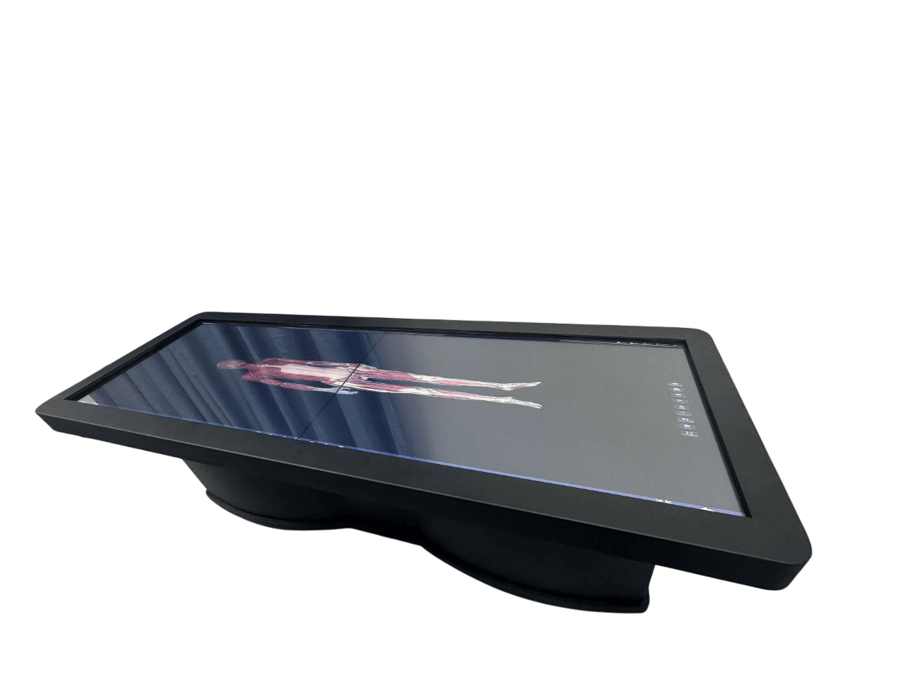
Systematic Anatomy : This module contains 3D structures obtained through the 3D reconstruction of real human cross-sectional data. The positions and shapes of these structures remain consistent with the original data. They are categorized into nine anatomical systems and can display the 3D morphology of over 6,000 anatomical structures.
Regional Anatomy : This module features peel and see-through functions to demonstrate structures from superficial to deep, facilitating the understanding of local hierarchical relationships and adjacent structures, even in a classroom setting. Additionally, it includes a large collection of regional anatomy teaching videos to support both teaching and independent student learning.
Sectional Anatomy : This module provides cross-sectional views of any body part. Using the highlighting function, students can quickly identify the Chinese and English names of anatomical structures, along with their positions and shapes in the 3D human body. It also includes real specimen images to enhance anatomical study.
Anatomy Micro Course : This module includes systematic anatomy, regional anatomy, and sectional anatomy micro-courses, with a total of over 130 courses.
Autonomous Learning : This module offers courseware composed of text, images, micro-videos, and 3D anatomical structures, serving as an essential learning resource for pre-class preparation and post-class review. The courseware also includes corresponding exercises, covering both theoretical and specimen-based assessments to support specimen examination. The module features over 1,800 exercises to reinforce knowledge retention.
Product Brochure Link here