S.M. Scientific Instruments (P) LTD

Home ≫Mortuary >Virtual Dissection Table and Digital Systems > Digital Dissection Table > Digital Virtual Anatomy Table SMI-DVAT

CLICK TO SCAN
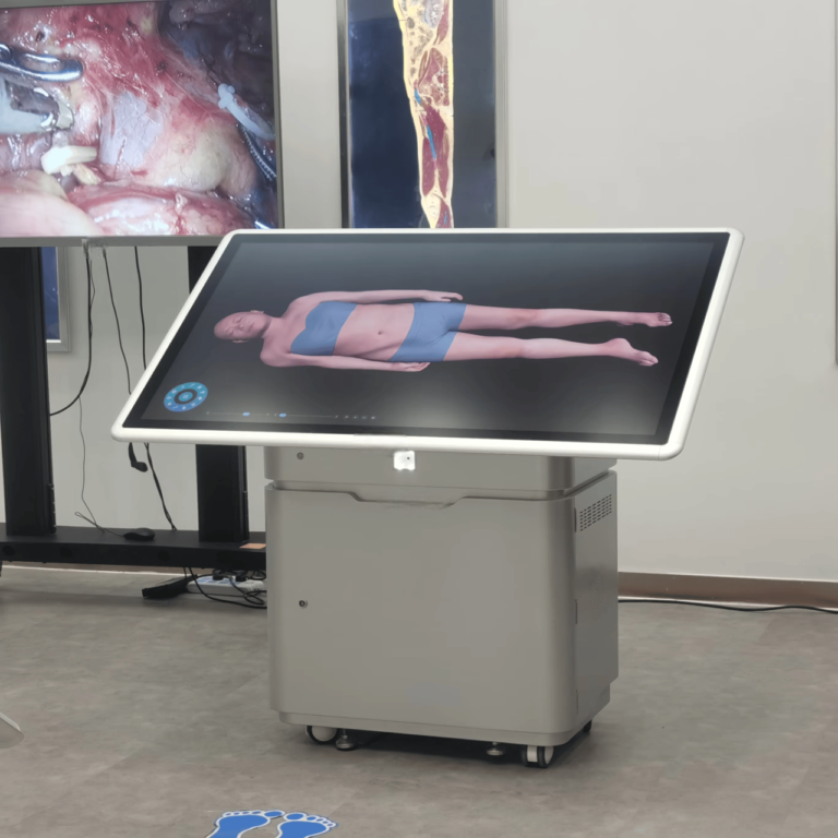
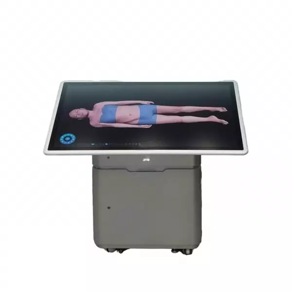
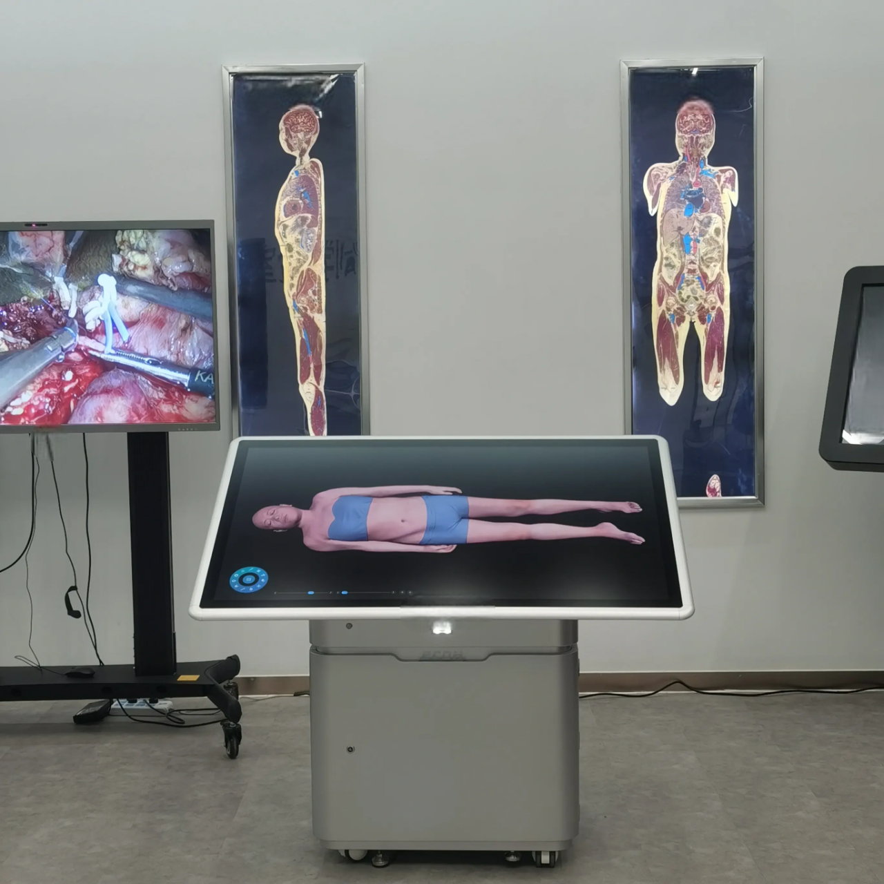
The HD Digital Virtual Anatomy Table uses real human body data reconstruction to simulate the actual anatomy process, combining traditional anatomy teaching with technology to provide an ideal learning solution. The screen can be lifted and tilted to accommodate different teaching scenarios.
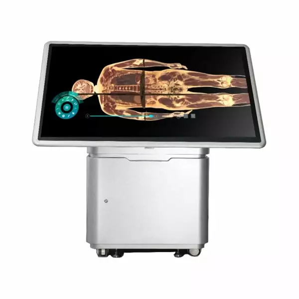
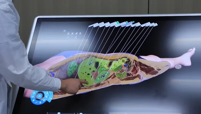
The Digital Virtual Anatomy Table is an advanced anatomical tool that creates a virtual representation of the human body using digital 3D reconstruction technology. Its touch control operation allows users to cut and observe human body structures from different directions, angles, and layers, integrating knowledge of human anatomy and sectional anatomy.
The system combines various medical visualization resources and human-computer interaction techniques. It integrates the UHD realistic anatomy system, the Digital Anatomy System, the user interaction management system, and other components to construct a comprehensive virtual training platform. With features such as a high-precision realistic anatomical structure, a high-performance anatomy teaching tool, and an interactive touch control interface, it enables users to explore the macroscopic and microscopic 3D structures of both normal and pathological anatomy.
Human Anatomy : The 3D structure of the human body in the human anatomy module is digitally reconstructed using male data (17,000+ total layers of cross-sections; resolution: 13,700 × 6,340) and female data (16,000+ total layers of cross-sections; resolution: 12,000 × 5,700). It enables users to perform virtual anatomical operations.
Anatomy system : The system offers a wealth of learning resources, including more than 3,000 3D structures, over 3,000 sectional images, more than 1,700 CT/MRI images, over 100 teaching videos, and more than 1,800 exercises. Through the Digital Anatomy Teaching System, teachers can operate professional anatomy teaching software using a touchpad. Students can explore stereoscopic human anatomy structures by wearing 3D glasses, immersing themselves in an open, autonomous, and interactive virtual environment. This facilitates an efficient, safe, and cost-effective teaching process.
Embryology : The system integrates videos, animations, 3D models, and courseware into a comprehensive teaching platform. It is categorized into three modules: early human embryogenesis (General), the development of human embryonic organ systems (Monographs), and congenital malformations. Each module includes structured teaching resources such as a preface, general videos, key knowledge points, and an exercise bank.
Slice Library : The Slice Library module contains at least 395 digital histology sections and 780 digital pathology sections. It supports touch and mouse controls to simulate microscope operations. Users can adjust magnification with one-click options for 4X, 10X, 20X, and 40X objective lenses, pan to modify the observation position, and quickly access previously viewed or favorited sections.
Clinical Cases : The Clinical Case module includes no fewer than 180 real clinical cases. Each case provides information such as the disease name, basic details, chief complaint, imaging findings, and diagnosis. The system allows manual adjustment of CT/MRI image window width and position based on different anatomical areas, enabling users to quickly and efficiently examine various imaging contents.
Product Brochure Link here