Constructing a multi-structured 3D digital anatomical model using high-precision digitized human body data. Utilizing a full-color, multi-material 3D printer and eco-friendly resin materials to produce 1:1 highly realistic anatomical models.
Digital Human Model
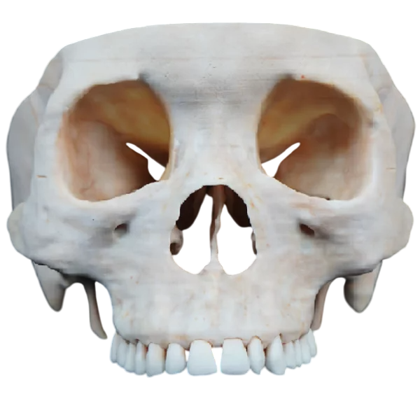
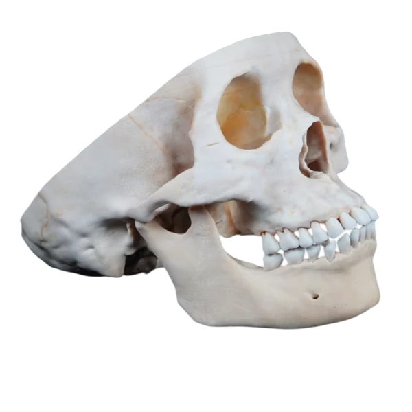
The skull base model represents the portion of the skull cut at the level of the complete skull, consistent with the skull top and mandible models of the same individual. It primarily displays the anatomical structure of the skull base, accurately restoring the anatomical features of the internal and external views of the skull base, as well as the front view of the skull. The displayed structures match the real bone specimen in terms of smoothness, roughness, and unevenness. The morphological features and visual details closely resemble those of actual bone specimens, making it an excellent substitute for skull specimens.
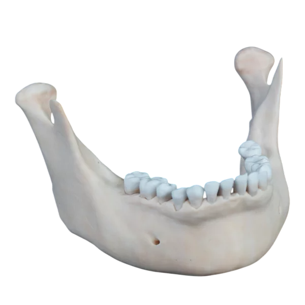
The mandible model is a complete, hard model of the mandible, including the full set of mandibular teeth, and is consistent with the skull base and top models. It accurately reflects the anatomical morphology and texture of the mandible, such as the smoothness of the mandibular ramus, the roughness of the mandibular mental bulge, and the concavities and convexities between the submandibular fossa and the mandibular hyoid muscle line. The model aligns with the bone specimens and serves as a suitable substitute for mandibular specimens.
Data Source: The data is selected from a high-precision digital human dataset, which includes original sectional data, refined segmentation data, and three-dimensional geometric models of organ structures such as bones, muscles, blood vessels, nerves, and ligaments. The voxel size of this dataset is 0.0384mm × 0.0384mm × 0.1mm. Additionally, cadaveric specimens fixed with formalin are used as a reference for comparison with the 3D-printed model.
Model Making: Voxels from the volume data on the surface of each anatomical structure are extracted from the original sectional dataset, and a texture map of the geometric model is generated to ensure that the appearance of the geometric model of each anatomical structure closely resembles that of the real anatomical specimen.

