S.M. Scientific Instruments (P) LTD

Home ≫Mortuary >Virtual Dissection Table and Digital Systems > Digital Anatomy System >Digital Anatomy Teaching System SMI-DATS

CLICK TO SCAN
The Digital Anatomy Teaching System integrates a vast amount of real human cross-sectional data into a computer to reconstruct a three-dimensional image of the human body. It is the result of a fusion of medicine, information technology, and computer technology. Currently, it is the only digital human anatomy product based on complete sectional reconstruction.
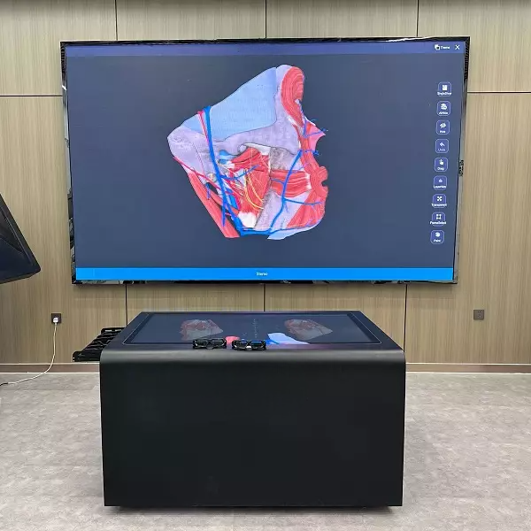
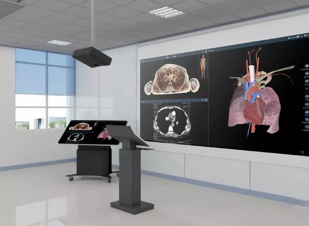
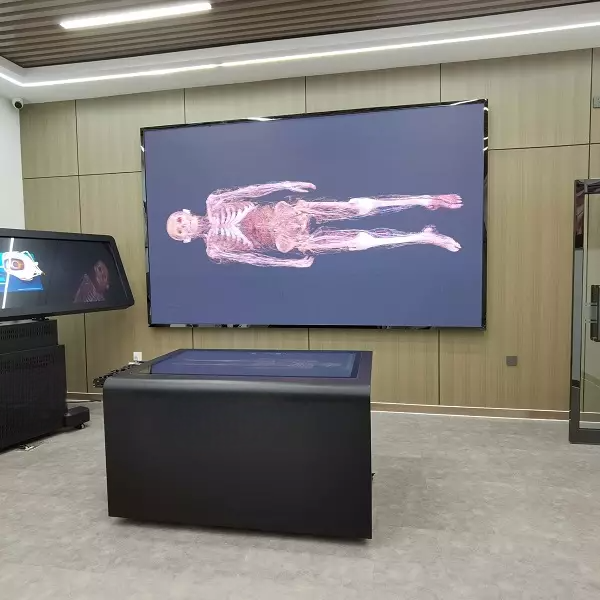
The system adopts an operation display mode that combines a touch podium with 3D projection and integrates the Chinese Digital Human Anatomy System. It utilizes advanced high-definition digital image processing and digital network technology while merging traditional anatomy experiment courses with cutting-edge computer virtual simulation technology. This approach provides students and doctors with an innovative learning environment, optimizes the teaching and training process, and enhances learning engagement through a novel digital anatomy curriculum and 3D display mode, ultimately improving teaching and training effectiveness.

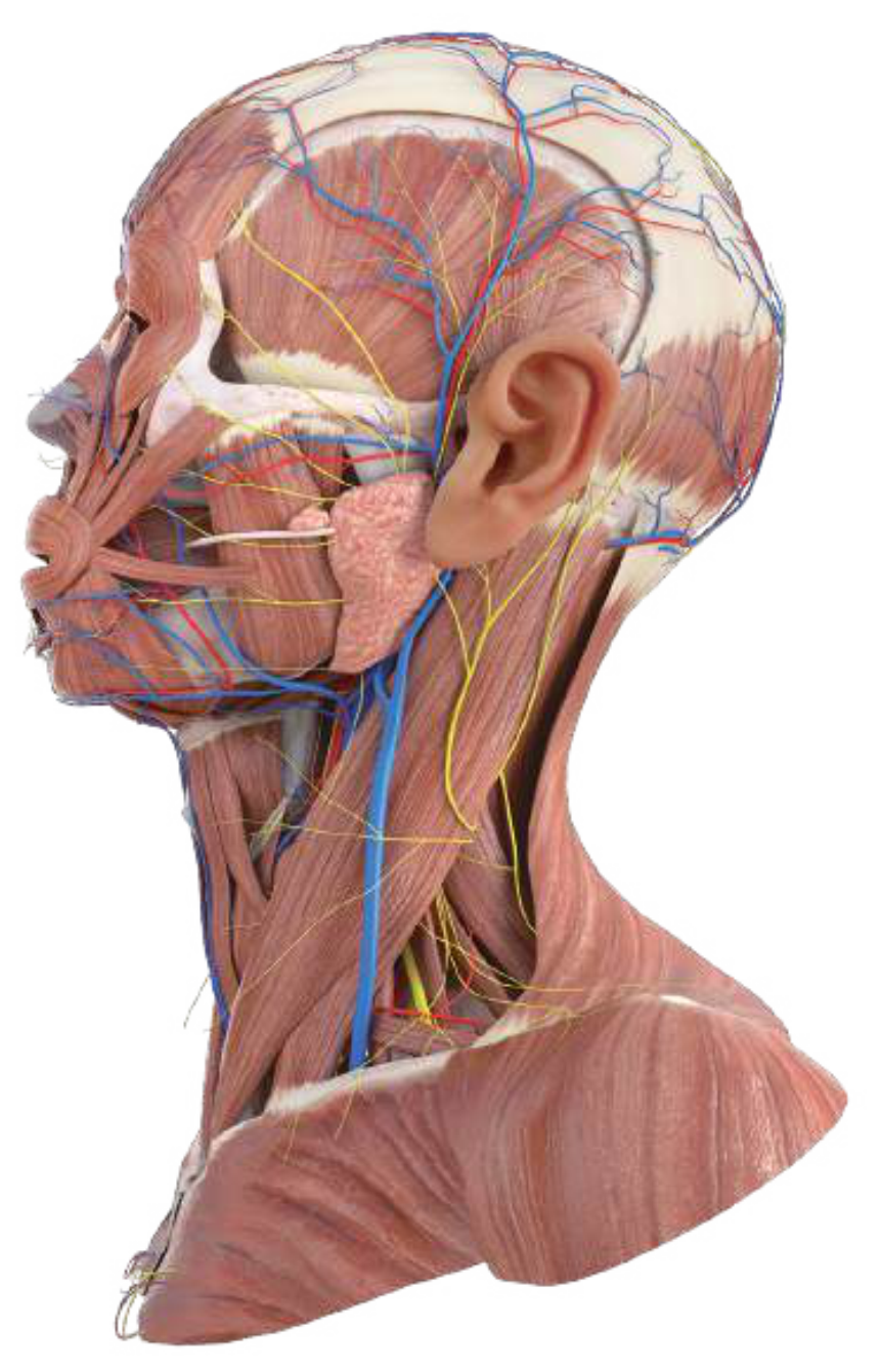
Professional digital human anatomy system : based on three-dimensional reconstruction of continuous real tomographic images, the digital human anatomy system uses real data from continuous tomography of a Chinese human body without organic diseases or defects. It reconstructs more than 6,000 anatomical structures in 3D with complete original data, including a normal appendix, normal teeth, and normal testes, without any missing segmental data. The digital human cross-sectional distances are as follows: head and neck ≤0.5mm (skull base ≤0.1mm), and other parts ≤1.0mm, with a total of over 2,100 tomographic data points. The system provides real human tomographic images in cross-sectional, coronal, and sagittal planes, which can be zoomed in and out freely, with a resolution of ≤0.18mm × 0.18mm per pixel.
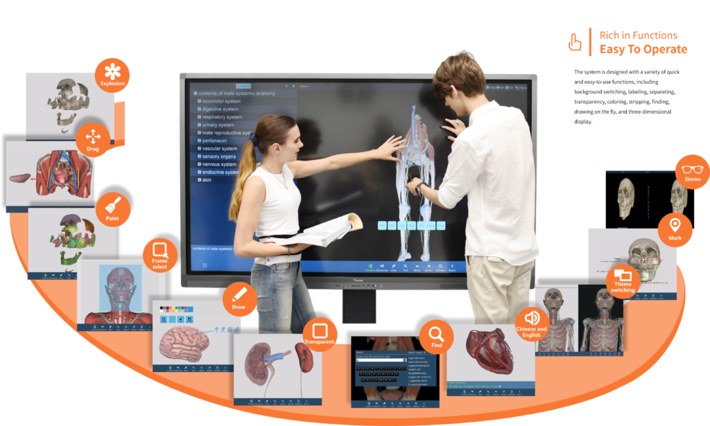
A fully functional digital anatomy teaching system : Following a structured catalog, all human organs and tissues are presented as true three-dimensional models, with key structure annotations and corresponding textual explanations. The models can be rotated and observed from any angle, including top-down and bottom-up perspectives. Users can add custom 3D annotations to each anatomical structure. Additional system features such as dissection, separation, and dyeing functions enhance the vividness, engagement, and intuitiveness of anatomy teaching.
Abundant teaching resources: The system encompasses systemic anatomy, regional anatomy, and tomographic anatomy. It includes over 1,700 corresponding CT and MRI images mapped to tomographic specimens, along with more than 130 system and local anatomical solutions. Additionally, it provides tomographic anatomy micro-lesson videos and a vast collection of digital exercises to enrich the learning experience.
Teaching Desk: 55-inch screen with a resolution of 3840×2160, brightness of 400 cd/m², and a capacitive 10-point touch system.
Computer: Equipped with an Intel Core i5 CPU, 8GB DDR memory, and a choice between a 240GB solid-state drive or a 500GB mechanical hard drive. Includes a wireless network card and a 2GB dedicated graphics card supporting 4K output.
Projector: Educational engineering projector with DLP projection technology, a nominal brightness of 5000 lumens, a standard resolution of 1920×1080 dpi, and a contrast ratio of 2000:1. Supports stereoscopic 3D technology projection and comes with two pairs of active stereo glasses as standard.
Product Brochure Link here