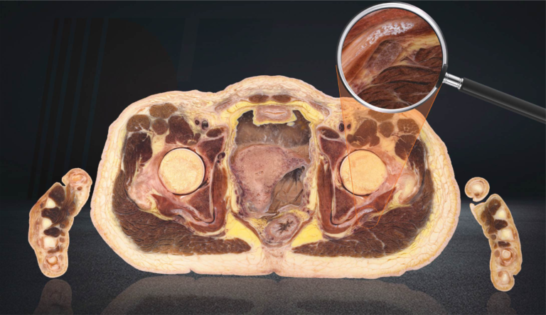
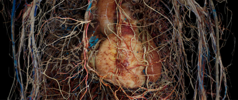
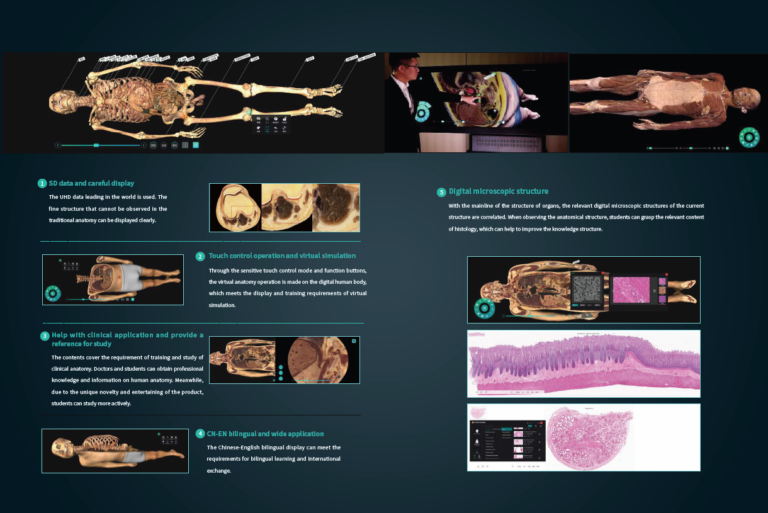
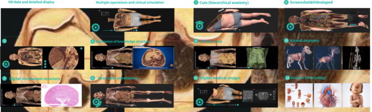
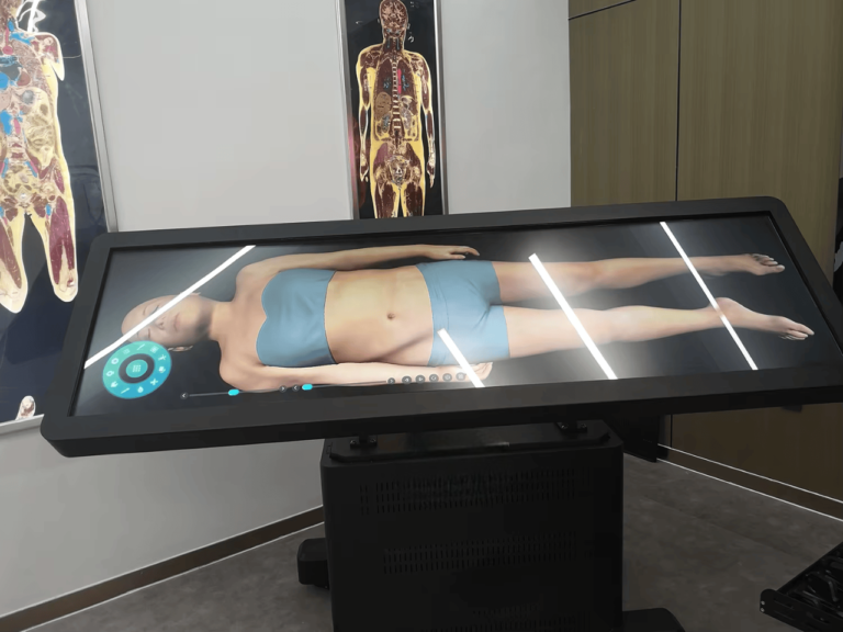
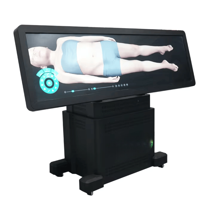
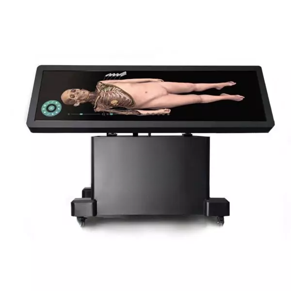
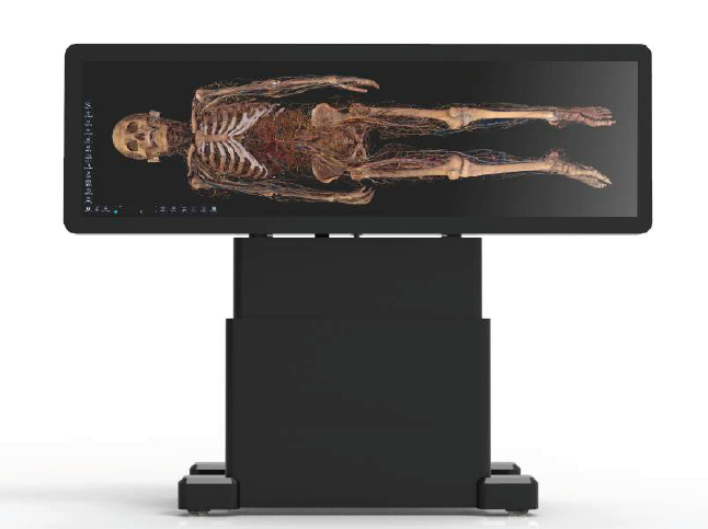
Human Anatomy : The 3D structure of the human body in the human anatomy module is digitally reconstructed from male data (17,000+ total cross-sectional layers; resolution: 13,700 × 6,340) and female data (16,000+ total cross-sectional layers; resolution: 12,000 × 5,700). Virtual anatomy operations can be performed using this module.
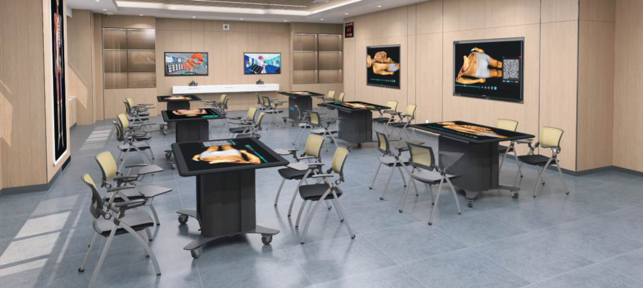
Anatomy System : The system contains a wealth of learning resources, including more than 3,000 3D structures, 3,000 sectional images, 1,700+ CT/MRI images, 100+ teaching videos, and 1,800+ exercises. Through the Digital Anatomy Teaching System, teachers can operate professional anatomy teaching software via the touchpad. Students can explore stereoscopic human anatomy structures using 3D glasses, allowing them to study in an open, autonomous, and interactive virtual environment. This enables an efficient, safe, and cost-effective teaching process.
Embryology : This system integrates videos, animations, 3D models, and courseware into a comprehensive teaching platform. It is categorized into three modules: General (early human embryogenesis), Monographs (development of human embryonic organ systems), and Congenital Malformations. Each module includes teaching resources such as a preface, general video, key knowledge points, and an exercise bank.
Slice Library : The slice library module contains at least 395 digital histology sections and 780 digital pathology sections. It supports touch or mouse controls to simulate microscope operations. Users can adjust magnification with one click (4X, 10X, 20X, 40X), pan to change the observation position, and access previously viewed or favorite sections.
Clinical Cases : The clinical case module includes no fewer than 180 real clinical cases. Each case displays the disease name, basic patient information, chief complaint, imaging findings, and diagnosis. The system allows manual adjustment of CT/MRI image window width and position according to different anatomical regions, enabling users to quickly analyze various image contents.

